
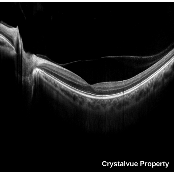
Vision 700 provides high resolution OCT images. It is also equipped with fundus camera.
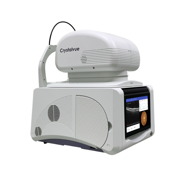
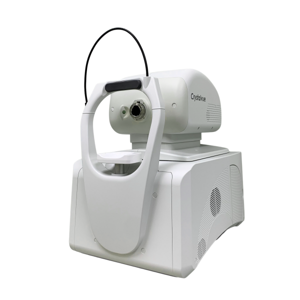
VISION-700
Features:
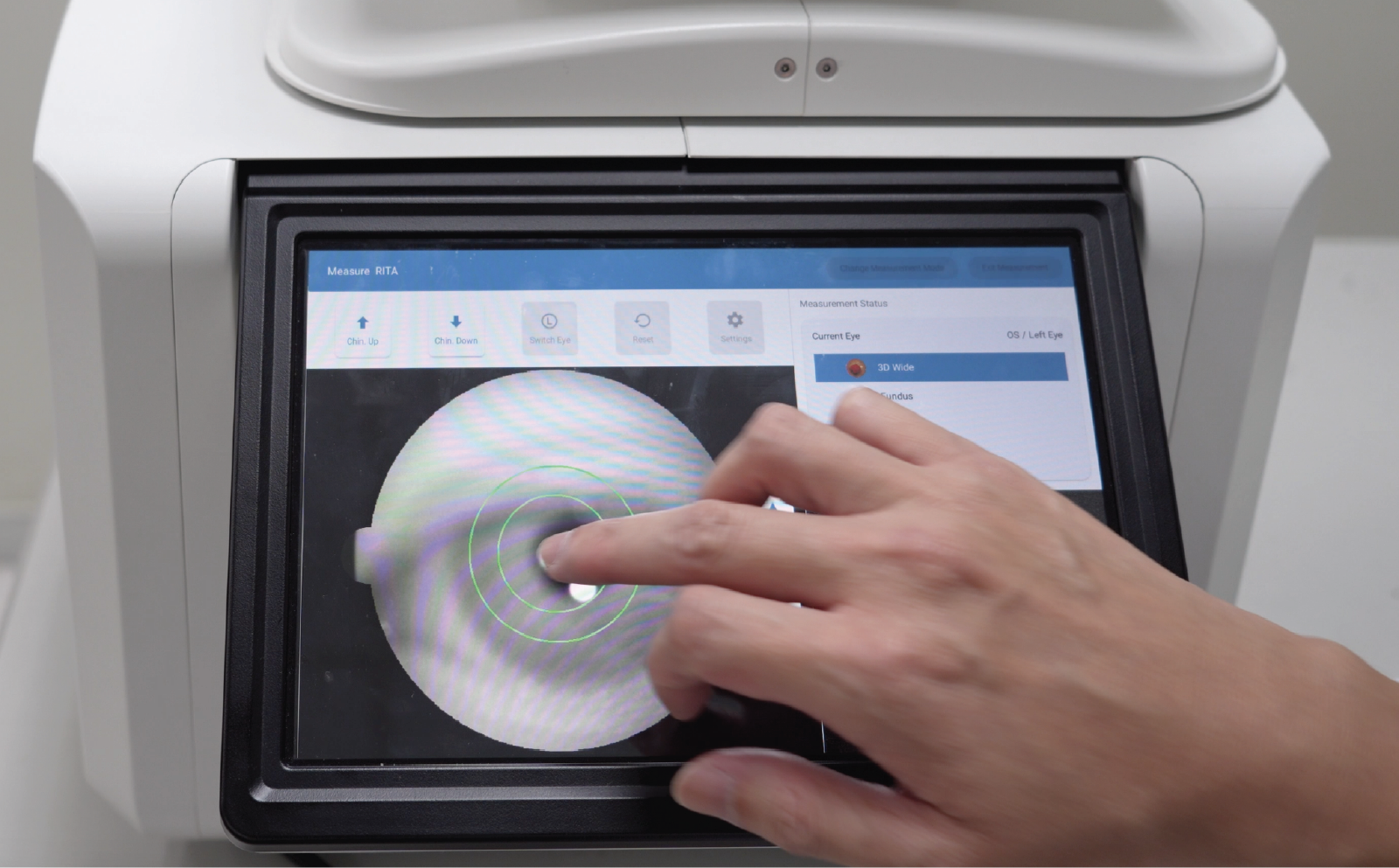
- 3D Auto Tracking
- Auto tracking and auto alignment by simply one tap on the pupil.
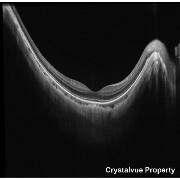
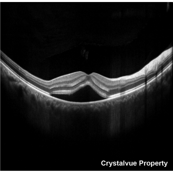
- High Speed High Resolution
- Generates high quality OCT image with high speed by 80,000 Hz A-Scan
- Fundus Camera Included
- Provides OCT, cornea images and also fundus photo.
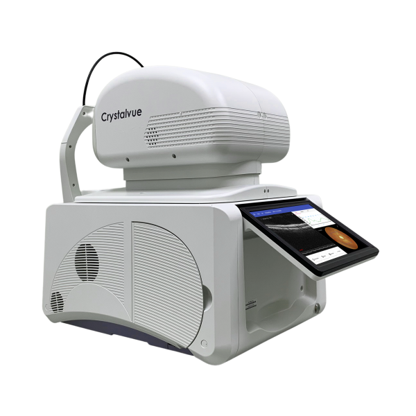
- Touch Screen
- Easy to operate. Able to zoom in and out to see the details.
Vision 700 provides high resolution OCT images. It is also equipped with fundus camera.
OCT and Fundus Camera Integrated
Crystalvue Vision 700 is a fully-automated, non-contact, high resolution tomographic and biomicroscopic imaging device. It also incorporates a non-mydriatic digital fundus camera and a built-in Windows 10 OS computer.
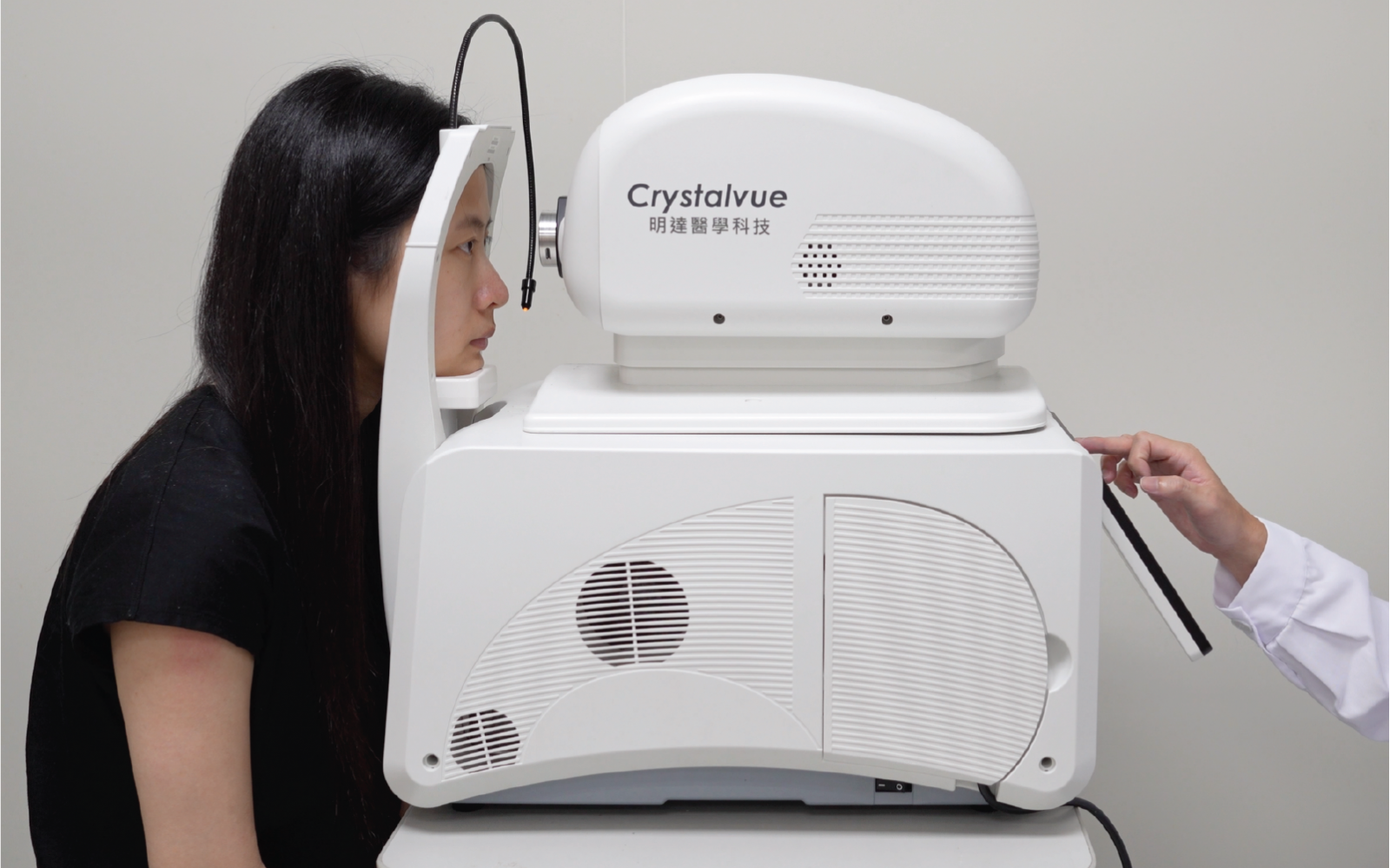
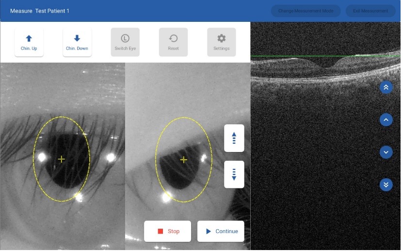
Fully Automatic Image Capture
Simply tap the pupil on the screen, Vision 700 will start tracking. With a single tap, Vision 700 can automatically align, focus, track, capture images and provide measurement results for Macula OCT and Disc OCT.
.png)
High Quality Images with 80k Hz A-Scan
Vision 700 captures and generates high-resolution OCT image and 12 MP high quality true color retinal photos.
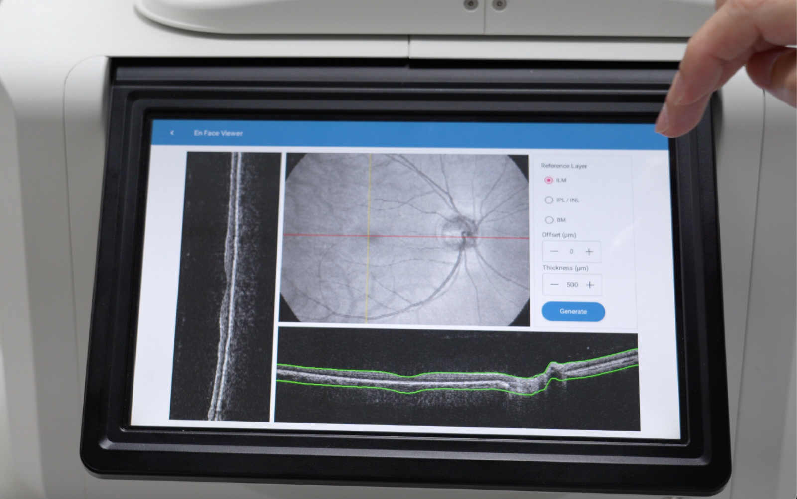
En Face Viewer
Vision 700 OCT/Fundus overlay feature can be applied to any designated area rather than merely the fovea.
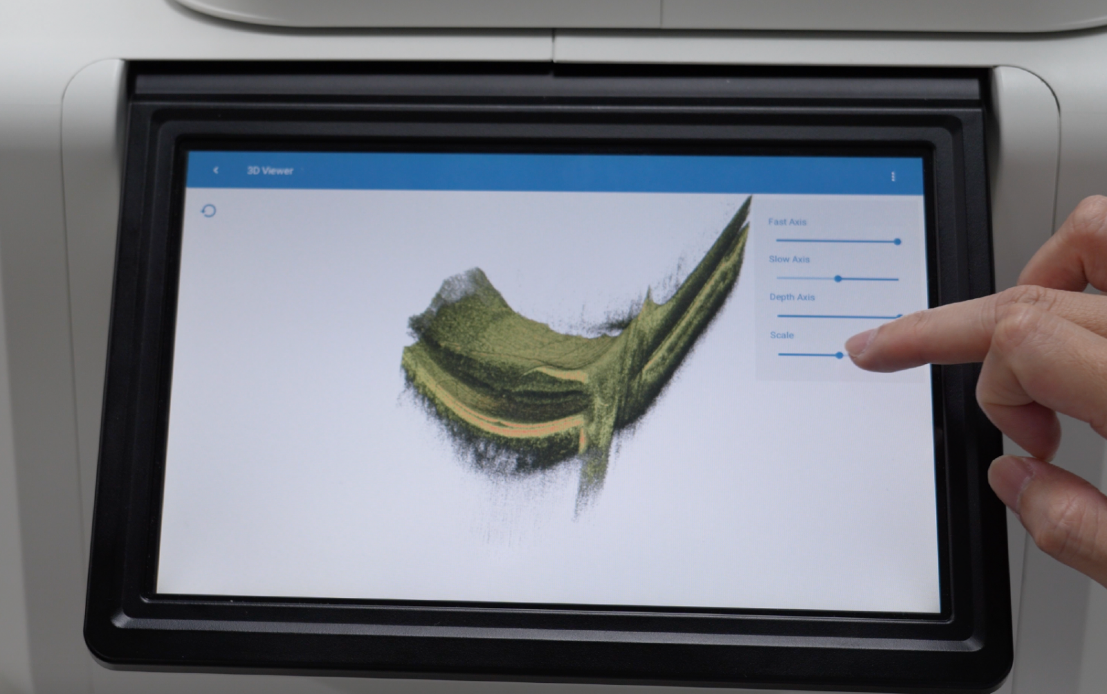
3D Viewer
Vision 700 is capable of reconstructing the retinal tissue in 3D space for inspection from a user-defined perspective and scale.
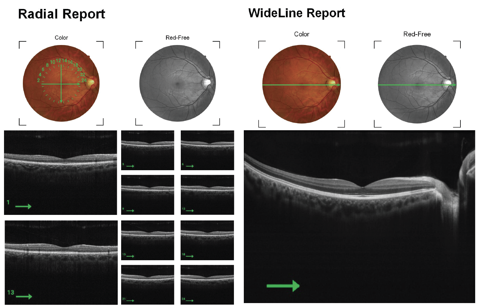
10 Scan Modes
Vision 700 provides measurement of posterior ocular structures, including retina, retinal nerve ber layer, macula and optic disc as well as imaging of anterior ocular structures. Users can select up to 10 Scan Modes.
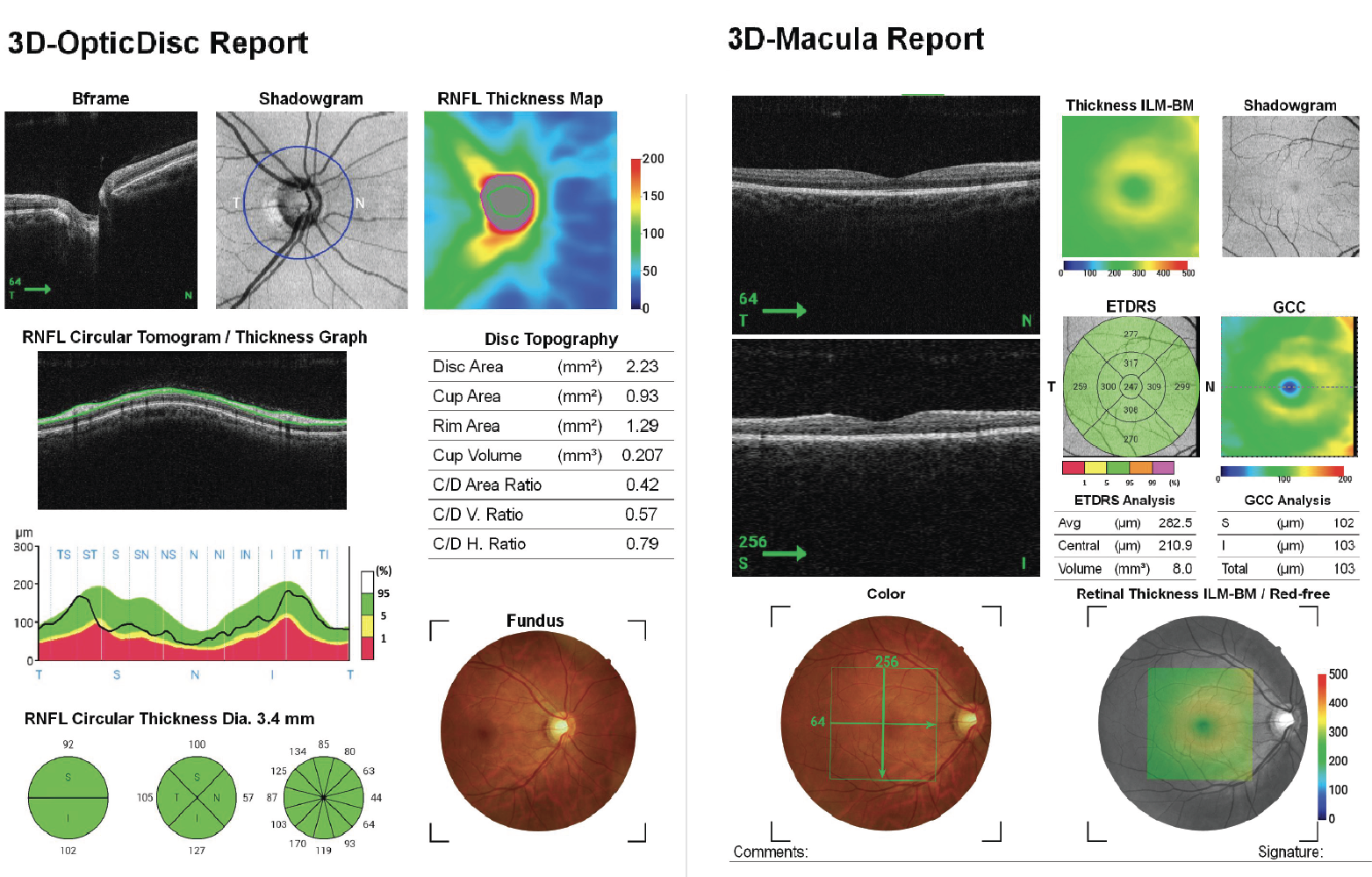
Various Comprehensive Analysis Reports
Vision 700 offers comprehensive analysis reports for each scan mode, including TSNIT, AVERAGE and ONH in 3D Optic Disc mode; THICKNESS, ETDRS and GCC in 3D Macula mode.
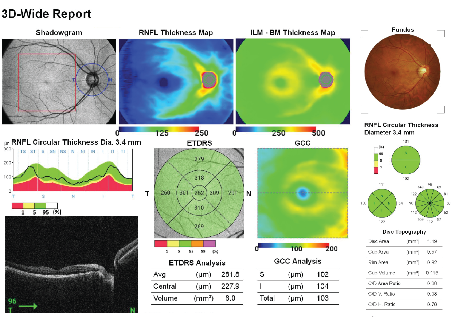
3D Wide Report
The wide scan range of 12×9 mm is supported.
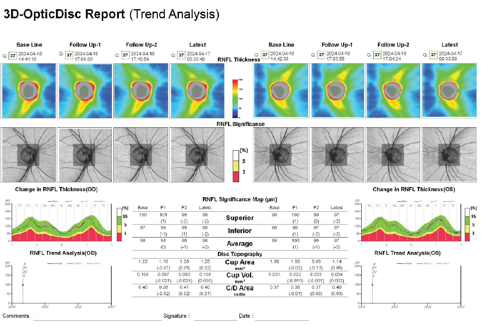
AI-Based Trend Analysis
Vision 700 provides AI-based trend analysis with up to four measures of both eyes in 3D Macula or 3D Optic Disc mode, giving clinicians more diagnostic options.
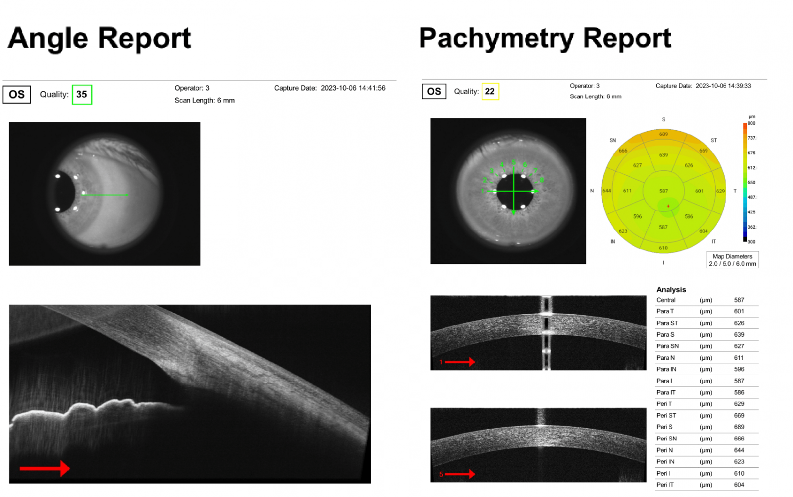
Pachymetry and Angle Mode
Vision 700 performs auto measurement for Pachymetry OCT and anterior ocular when the CAM (optional accessory) is attached.
Manual Mode
Besides Auto mode, Vision 700 also supports Manual mode for OCT measurements.
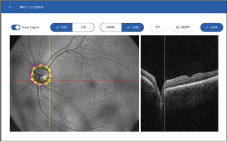
Built-in Photo Edit Functions
Users are able to edit disc, cup border, and layers to reanalyze the result. There are also multiple filters including RGB, negative film, and red-free for users to apply.
Specification
| Model Name: VISION 700 | |
|---|---|
| Functionality | Automated Optical Coherence Tomography (OCT), Fundus Camera (FC) |
| Computer | Integrated with the device. Window 10 IoT Enterprise version |
| Display | 10.1in LCD with Touch function, 1280×800 pixel |
| Alignment | Automatic 3D Tracking/Focusing, Manual |
| Light Source | Optical Coherence Tomography: SLED 840nm |
| Scan Mode | 3D Mode: 3D Optic Disc, 3D Macula / Line Mode: Line, Wide Line, 5-line Cross, Radial |
| Scan Mode | Pachymetry and Angle Measurement (Anterior Chamber) |
| Scan Range | Line and 3D Mode: 6mm x 6mm (H & V ± 5%), Wide Line Mode: 12mm or less (± 5%) |
| Scan Speed | 80KHz A-scan |
| In-depth Resolution | < 6μm |
| Fixation Target | 15 points internal (Green), 1 adjustable external (Amber) |
| Type of Photograph Review | True Color, Red-free, Negative Film |
| Field of Angle | 45 °± 5% |
| Focus Adjustment Range without compensation lens | -15D to +10D |
| Focus Adjustment Range with compensation lens | -30D to -10D, +5D to +30D |
| Photographable Pupil Size | ø2.5mm or more via OCT; ø3.8mm or more via FC |
| Fundus Image Resolution | 12M Pixel |
| Interface of Connectivity | HDMI, USB3.0 (blue), USB2.0 (white), RJ45/Ethernet |
| OM Operating Range | Front/Back 65mm, Left/Right 100mm, Up/Down 30mm |
| Chinrest Adjustment Range | Up/Down 70mm |
| Power Supply | Medical Grade, AC100-240V@50-60Hz, Auto. Power Consumption < 400VA |
| Dimension / Weight | W 409mm, D 534mm, H 546mm / 32kg |